Home » Without Label » Labeled Diagram Of An : Anatomy Bone Labeling Worksheet Sheets | Skeletal system ... - All these parts combine and work together.
Labeled Diagram Of An : Anatomy Bone Labeling Worksheet Sheets | Skeletal system ... - All these parts combine and work together.
Labeled Diagram Of An : Anatomy Bone Labeling Worksheet Sheets | Skeletal system ... - All these parts combine and work together.. Who let the hulk out? Learn vocabulary, terms, and more with flashcards, games, and other study tools. Labeled diagram of the human kidney. All these parts combine and work together. Posted on january 16, 2016 by admin.
You should now be able to label all the main anatomical features. The primary parts of anatropous (ovule) are: Find free pictures, photos, diagrams, images and information related to the human body right here at science kids. (iii)micropyle is an opening presentation at the tip where integument is missing. This diagram depicts labeled muscle diagram 1024×1878 with parts and labels.
Volcano diagrams from static.wixstatic.com (iv) chalaza is inverse to the micropylar end speaking to the basal piece of the ovule. We think this is the most useful. There are different ways of dividing the brain anatomically into regions. Let learn the different parts of the human digestive system. The anatomy of the knee consists of bones, muscles, nerves, cartilages, tendons and ligaments. Labeled diagram of the knee joint. The structure of an atom explained with a labeled diagram. For more anatomy content please follow us and visit our website:
Here is the labeled diagram.
The anatomy of the knee consists of bones, muscles, nerves, cartilages, tendons and ligaments. Human eye anatomy, retina, optic disc artery and vein etc. It is the beginning of the digestive tract and the process of digestion begins from the mouth, where teeth help by breaking and grinding the food. Human organs & anatomy diagram picture category: These bones are arranged into two major divisions: Their observations led to the discovery of sarcomere zones. Labelled diagram drag and drop the pins to their correct place on the image. A neuron is a specialized cell, primarily involved in transmitting information through electrical and chemical signals. The axial skeleton runs along the body's midline axis and is made up of 80 bones in the following regions: The muscles of the lower back help stabilize, rotate, flex, and extend the spinal column, which is a bony tower of 24 vertebrae that gives the body structure and houses the spinal cord. Knee joint is one of the most important hinge joints of our body. By the end of this post, you will be able to label the anatomical features shown on the diagram below. There are different ways of dividing the brain anatomically into regions.
Posted on january 16, 2016 by admin. (i) the hilum is an intersection among ovule and funicle. We hope this picture labelled diagram of the muscles in the human body can help you study and research. The anatomy of the knee consists of bones, muscles, nerves, cartilages, tendons and ligaments. Labeled diagrams can be used in math, science and language arts in order to help students understand the relationships among different parts of the diagram.
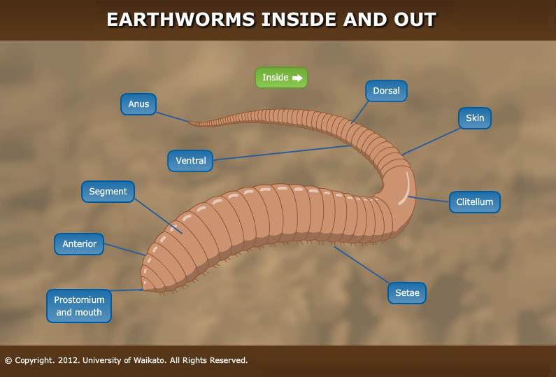
Outside of an earthworm — Science Learning Hub from www.sciencelearn.org.nz Their observations led to the discovery of sarcomere zones. Find free pictures, photos, diagrams, images and information related to the human body right here at science kids. A labeled diagram is a drawing, chart or graph that is used to visualize a concept. 674 x 599 photo description: Brain diagram with labels hypothalamus vector brain diagram pons cerebrum and cerebellum brain pons brain anatomy amygdala brain labelled amygdala brain human midbrain diagram pons. These bones are arranged into two major divisions: The following article provides you with diagrams that will help you understand the structure of an atom better. Learn vocabulary, terms, and more with flashcards, games, and other study tools.
This diagram depicts labeled muscle diagram 1024×1878 with parts and labels.
Find free pictures, photos, diagrams, images and information related to the human body right here at science kids. Find a great range of the diagram of human body and anatomy diagrams in the following pictures. The system breaks down food, extracts nutrients from it, and converts them into energy. The human digestive system is the means by which tissues and organs receive nutrients to function. Brain diagram with labels hypothalamus vector brain diagram pons cerebrum and cerebellum brain pons brain anatomy amygdala brain labelled amygdala brain human midbrain diagram pons. The heart is a muscular organ about the size of a fist, located just behind and slightly left of the breastbone. A neuron is also known as the nerve cell. The axial skeleton runs along the body's midline axis and is made up of 80 bones in the following regions: (iii)micropyle is an opening presentation at the tip where integument is missing. Labelled diagram drag and drop the pins to their correct place on the image. Utilize the model of the human brain to locate the following structures / landmarks for the By the end of this post, you will be able to label the anatomical features shown on the diagram below. The muscles of the lower back help stabilize, rotate, flex, and extend the spinal column, which is a bony tower of 24 vertebrae that gives the body structure and houses the spinal cord.
The following article provides you with diagrams that will help you understand the structure of an atom better. The anatomy of the femur can be divided into proximal, central, distal, and posterior parts. All these parts combine and work together. Diabetes and healthy eyes toolkit and website keywords: Label fractions on number line.
Osmosis diagram from cdn.thinglink.me Diabetes and healthy eyes toolkit and website keywords: Learn vocabulary, terms, and more with flashcards, games, and other study tools. You should now be able to label all the main anatomical features. We think this is the most useful. These bones are arranged into two major divisions: A neuron is a specialized cell, primarily involved in transmitting information through electrical and chemical signals. Let's use a common method and divide the brain into three main regions based on embryonic development: The following article provides you with diagrams that will help you understand the structure of an atom better.
Learn vocabulary, terms, and more with flashcards, games, and other study tools.
The human kidneys house millions of tiny filtration units called nephrons, which enable our body to retain the vital nutrients, and excrete the unwanted or excess molecules as well as metabolic wastes from the body. The axial skeleton and the appendicular skeleton. The forebrain, midbrain and hindbrain. In addition, they also play an important role in maintaining the water balance of our body. Mitosis is a process of cell division which results in the production of two daughter cells from a single parent cell. The anatomy of the femur can be divided into proximal, central, distal, and posterior parts. The structure of a neuron varies with their shape and size and it mainly depends upon their. The heart pumps blood through the network of arteries and. Let learn the different parts of the human digestive system. This diagram of the human body shows a range of organs that are important to human anatomy.they include the brain, heart, lungs, spleen, muscles. The primary parts of anatropous (ovule) are: The human digestive system is the means by which tissues and organs receive nutrients to function. Let's use a common method and divide the brain into three main regions based on embryonic development: